New Imaging Technique Could Transform Precision of Vocal Fold Injection Procedures
Shortwave infrared imaging offers a new path to better patient outcomes.
AAO-HNSF
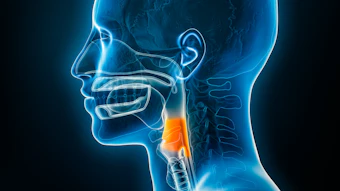
Researchers at Stanford University, in collaboration with scientists at the German Cancer Institute, have shown for the first time that shortwave infrared (SWIR) imaging can be used to visualize injectable filler materials during injection laryngoplasty, a common procedure used to treat vocal fold paralysis and other forms of glottic insufficiency. The findings, published in Otolaryngology–Head and Neck Surgery, on this novel visualization technique could pave the way for precision-guided laryngeal surgery.
 Tulio A. Valdez, MD, MSc
 Roy Kiwan Park, MD
The combination of reflection and fluorescence SWIR imaging offers a multimodal approach that could help surgeons confirm correct filler placement in real time, identify misplaced superficial injections, and potentially monitor filler resorption over time.
While the study was conducted in a laboratory setting using ex vivo tissues, the authors note that the optical setups could be integrated into endoscopes or operating microscopes for clinical use. The cost of SWIR camera technology has decreased substantially in recent years, making broader clinical adoption increasingly feasible.
Beyond injection laryngoplasty, the researchers suggest SWIR imaging could have future applications in identifying vocal fold pathologies such as cysts and polyps, differentiating layers of the vocal fold based on water and collagen content, and detecting areas of laryngeal inflammation.
Reference
Park, R.K., Lee, M.C., Härtl, S., Arús, B.A., Nuyen, B., Sung, C.-K., Baik, F.M., Bruns, O.T., and Valdez, T.A. (2026). Multimodal Shortwave Infrared Imaging for Visualization of Injection Laryngoplasty. Otolaryngol Head Neck Surg, 174: 185-194. https://doi.org/10.1002/ohn.70050