Revisiting Max Brödel’s 1939 Classic Coronal Illustration of the Ear
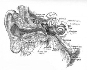
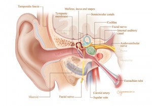
Robert K. Jackler, MD, Christine Gralapp, Albert Mudry, MD, PhD, for the Otolaryngology Historical Society In 1939, the renowned Johns Hopkins medical artist Max Brödel created a coronal illustration of the ear (figure 1). Brödel’s magnificent and hugely successful illustration drew upon artistic conventions developed during the preceding 200 years. It nevertheless constituted an important improvement over prior depictions in large part due to his consummate skill as an illustrator. As a schematic, it provided clarity to complex anatomical relationships in a manner that was readily understandable. For nearly seven decades, Brödel’s magnificent illustration has served as the inspiration for innumerable textbook and article illustrations. In his design, the artist intentionally choose to diverge from literal anatomy in that he distorted some structures (such as rotating the incus 180 degrees, bending the cochlea anteriorly, and substantially enlarging the inner ear) to bring them into greater prominence and clarity and eliminated others (such as the carotid artery and intratemporal facial nerve) to avoid a cluttered image. Several anatomical errors exist, such as the absence of the scutum and a markedly foreshortened internal auditory can Brödel’s illustration has been routinely imitated by subsequent illustrators (in collaboration with otologists) and virtually all have faithfully reproduced Brödel’s purposeful artistic distortions and unintentional errors in their depictions—often with the assumption that they represented actual anatomy rather than an artistic interpretation. Perpetuation of distortions and errors in anatomical illustration is not a rare phenomenon. This led us to offer a more anatomically accurate standard coronal schematic of the ear, created by artist Christine Gralapp in collaboration with Drs. Robert Jackler and Albert Mudry, which we hope will enhance the clarity and precision of future illustrations in the otological literature (figure 2).
 Figure 1. Max Brödel’s 1939 Classic Coronal Illustration of the Ear (now in the public domain).
Robert K. Jackler, MD, Christine Gralapp, Albert Mudry, MD, PhD, for the Otolaryngology Historical Society
In 1939, the renowned Johns Hopkins medical artist Max Brödel created a coronal illustration of the ear (figure 1). Brödel’s magnificent and hugely successful illustration drew upon artistic conventions developed during the preceding 200 years. It nevertheless constituted an important improvement over prior depictions in large part due to his consummate skill as an illustrator. As a schematic, it provided clarity to complex anatomical relationships in a manner that was readily understandable.
For nearly seven decades, Brödel’s magnificent illustration has served as the inspiration for innumerable textbook and article illustrations. In his design, the artist intentionally choose to diverge from literal anatomy in that he distorted some structures (such as rotating the incus 180 degrees, bending the cochlea anteriorly, and substantially enlarging the inner ear) to bring them into greater prominence and clarity and eliminated others (such as the carotid artery and intratemporal facial nerve) to avoid a cluttered image. Several anatomical errors exist, such as the absence of the scutum and a markedly foreshortened internal auditory can
 Figure 2. The comparison illustration is reproduced by courtesy of Stanford University
Brödel’s illustration has been routinely imitated by subsequent illustrators (in collaboration with otologists) and virtually all have faithfully reproduced Brödel’s purposeful artistic distortions and unintentional errors in their depictions—often with the assumption that they represented actual anatomy rather than an artistic interpretation. Perpetuation of distortions and errors in anatomical illustration is not a rare phenomenon. This led us to offer a more anatomically accurate standard coronal schematic of the ear, created by artist Christine Gralapp in collaboration with Drs. Robert Jackler and Albert Mudry, which we hope will enhance the clarity and precision of future illustrations in the otological literature (figure 2).


