Cerebrospinal Fluid Leaks: A Guide for the Comprehensive Otolaryngologist
CSF leaks can lead to serious complications if untreated, underscoring the need for prompt diagnosis, risk assessment, and appropriate management.
Eric Nisenbaum MD, MSc, Shawn M. Stevens, MD, Corinna G. Levine, MD, MPH, on behalf of the Skull Base Surgery Committee

CSF leaks can be categorized by their etiology—either traumatic or nontraumatic. Approximately 80%-90% of cranial CSF leaks are secondary to trauma. This can be further subdivided into accidental trauma, such as a car crash or assault, and iatrogenic trauma after temporal bone or sinus surgery. The incidence of leaks after accidental trauma ranges from 2% (all closed-head injuries) to 30% if a skull base fracture is present. Leaks generally present early after the inciting event, with >50% starting within the first 48 hours after injury and 70% presenting within the first week.
Nontraumatic leaks, also referred to as spontaneous CSF leaks, comprise the other 10%-20% of cases. These are more common in the fourth to sixth decades of life and are seen more frequently in female than male patients. Nontraumatic leaks are often associated with elevated intracranial pressures (ICP), though a subset of patients may have normal ICP. Most patients with spontaneous CSF leaks have an overweight or obese habitus as defined by having a body mass index (BMI) >25-30kg/m.2 Approximately 70% of non-traumatic leaks are associated with idiopathic intracranial hypertension (IIH)—also known as pseudotumor cerebri—a condition in which ICP is elevated without clear underlying pathology.
Presentation
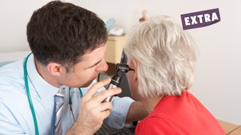
Patient presentation can vary based on the location and cause of the leak. Leaks from the lateral skull base can be associated with aural fullness and hearing loss due to CSF filling the middle ear space. Some patients may also report pulse-synchronous tinnitus, headaches, and mild dizziness. The latter is especially common in simultaneous presentations of IIH. In most cases, otoscopy will reveal clear fluid or possibly an air-fluid meniscus behind an intact tympanic membrane. Persistent, clear otorrhea may also be encountered in patients with a tympanic membrane perforation or a tympanostomy tube. In patients undergoing lateral skull base surgery, the leak can present as a fluctuant subcutaneous collection at the surgical site (also called a pseudomeningocele) or as leakage through the incision line.
Anterior skull base leaks are associated with clear rhinorrhea, which may be constant or intermittent. Presentations are generally unilateral. The rhinorrhea may be triggered or worsened by straining, lifting, or bending over. It can also present with post-nasal drip that has a salty and/or metallic taste. Notably, some lateral skull base leaks may also present with rhinorrhea or post-nasal drip as the primary complaint. This is due to the drainage of CSF through the eustachian tubes. Anosmia may be seen in patients with leaks at the cribriform plate associated with meningocele or neoplasm. Headaches are very common regardless of location or etiology, secondary to either over-drainage of CSF or elevated ICP. Patients should be screened for vision changes (blurring, loss of color, field restriction) as this may occur in patients with IIH over time. Patients with undiagnosed leaks may also initially present with meningitis.
Risk Factors
Although many of the signs and symptoms associated with CSF leak are nonspecific, some presentations are associated with an increased risk of having a leak or later developing leak-related complications. These include a history of facial or head trauma, recent sinonasal/neurotologic/cranial surgery, presence of sinonasal or intracranial tumor, chronic ear or sinus pathologies such as cholesteatoma, allergic fungal sinusitis, venous sinus stenosis, obesity, and obstructive sleep apnea (OSA). It is important to note that OSA raises CSF pressure due to high central venous pressures (high thoracic obstructive pressures), which transmit up to the dural venous sinuses, and which ultimately reduce CSF absorption via the arachnoid granulation system.
In patients with the above risk factors, a high index of suspicion for CSF leak should be maintained and the provider should have a lower threshold to obtain further diagnostic workup. Similarly, all patients with presentations that are concerning for a leak should also be screened for IIH symptoms.
Work-up
The work-up of a potential CSF leak starts with a detailed history and physical exam. Although many patients only leak intermittently, active leaking can sometimes be elicited by having a patient hang their head forward or with a Valsalva maneuver.
For patients with rhinorrhea or other sinonasal symptoms, an endonasal fiberoptic scope exam should be performed to evaluate for associated pathology such as an encephalocele or a tumor. The scope exam can also help localize the leak, though in non-operated sinuses, it may be difficult to visualize the exact location of the leak. In patients with otologic symptoms, in addition to a detailed examination, an audiogram with tympanometry should be performed. In such cases, a conductive or mixed hearing loss would be expected with a type B (flat) normal volume tympanogram if the tympanic membrane is intact.
To help identify a CSF leak, leaking fluid can be collected and tested for the presence of beta-2 transferrin, a protein only found in CSF, perilymph, and the aqueous/vitreous humors of the eye. In cases of CSF otorrhea, a tympanostomy can be performed to aid in the collection and testing of fluid. It should be noted that such testing requires at least 1 ml of fluid, ideally collected in a small, closed container that is refrigerated as soon as possible. This is because beta-2 transferrin protein will denature within hours at room temperature and thus produce a false negative result. Providers should also confirm whether their local lab facilities can perform such testing.
Imaging also plays a key role in the evaluation of a potential CSF leak. Both a CT with thin collimations (either of the sinuses or temporal bones depending on the suspected location) and an MRI of the skull base with and without contrast should be obtained as part of the imaging work-up, as they provide complementary information. The CT allows for a detailed evaluation of the bony anatomy to assess for skull base defects. Common areas of dehiscence include the cribriform plate, ethmoid and sphenoid roofs, and the tegmen. MRI provides a detailed evaluation of the soft tissue anatomy and is useful for identifying neoplasms and other soft tissue pathology such as meningoceles, encephaloceles, and cholesteatoma. MRI can also identify signs of elevated ICP/IIH such as an empty sella, flattening of the posterior sclera, tortuosity of the optic nerves with widening of the surrounding subarachnoid space, and venous stenosis.
For patients with small or multiple skull base defects, more specialized imaging modalities such as MR or CT cisternography can also be used to better pinpoint the leak. For patients with nontraumatic leaks, a lumbar puncture can help confirm the diagnosis of IIH. An opening pressure greater than 25 centimeters of water is consistent with IIH; however, it is important to remember that pressure may be spuriously lower during an active CSF leak which acts as a release valve for the increased pressure.
Treatment
Once a CSF leak has been diagnosed, management in part depends on the etiology of the leak. Traumatic leaks—either accidental or iatrogenic—are comparatively much more likely to resolve with conservative management (>90%). First-line therapy includes activity restriction—with avoidance of heavy lifting, straining, nose blowing, and other activities that raise ICP—as well as the use of a carbonic anhydrase inhibitor such as acetazolamide to decrease CSF production. The goal of these interventions is to decrease pressure gradients at the site of the leak to promote healing. If these interventions fail, it is an option to refer patients to neurosurgery for a high-volume lumbar puncture or lumbar drain placement to further reduce ICP. Otherwise, for non-resolving leaks a specialist referral should be made—either to rhinology or neurotology/otology, depending on leak location—for surgical repair. While the details of surgical repair are beyond the scope of this article, the goal is to re-establish separation between the intracranial and extracranial compartments while preserving function and minimizing dead space at the repair site.
Comparatively, non-traumatic leaks are less likely to heal spontaneously (<10-20%) and frequently require surgical repair. To maximize the chance of a successful repair, the underlying pathology—most commonly elevated ICP—must also be managed. Patients can be started on acetazolamide, counseled on the importance of weight loss if obese, and referred to neurology for further management. This is especially the case in suspected presentations of IIH. Patients should also be referred to neuro-ophthalmology for evaluation, with close follow-up if papilledema is identified. Other referrals might also be appropriate depending on underlying risk factors, including sleep medicine in patients with uncontrolled sleep apnea and endocrinology (there is a frequent association between many endocrine diseases and IIH). If IIH cannot be controlled with medical management and lifestyle changes, more invasive interventions such as ventriculoperitoneal shunting or venous sinus stenting may be necessary. Such interventions may also be necessary in cases of surgically refractory, recurrent leaks.
Summary
Patients with CSF leaks can present with a variety of common otolaryngology complaints, such as rhinorrhea or aural fullness, and thus are frequently first evaluated by comprehensive otolaryngologists. A high degree of suspicion and a thorough history and physical exam are crucial to identifying a leak. Beta-2 transferrin testing can confirm a leak, and imaging work-up with both CT and MRI is important to localize the leak and identify the underlying pathology. Classification into traumatic and nontraumatic leaks has important implications for prognosis and management. Providers should have a low threshold for specialist referral for leak repair and management of comorbid conditions.
References
- Georgalas C, Oostra A, Ahmed S, Castelnuovo P, Dallan I, van Furth W, et al. International Consensus Statement: Spontaneous Cerebrospinal Fluid Rhinorrhea. Int Forum Allergy Rhinol. 2021;11(4):794-803.
- Naples JG, Shah RR, Ruckenstein MJ. The evolution of presenting signs and symptoms of lateral skull base cerebrospinal fluid leaks. Curr Opin Otolaryngol Head Neck Surg. 2019;27(5):344-8.
- Oakley GM, Alt JA, Schlosser RJ, Harvey RJ, Orlandi RR. Diagnosis of cerebrospinal fluid rhinorrhea: an evidence-based review with recommendations. Int Forum Allergy Rhinol. 2016;6(1):8-16.
- Oh JW, Kim SH, Whang K. Traumatic Cerebrospinal Fluid Leak: Diagnosis and Management. Korean J Neurotrauma. 2017;13(2):63-7.
- Prosser JD, Vender JR, Solares CA. Traumatic cerebrospinal fluid leaks. Otolaryngol Clin North Am. 2011;44(4):857-73, vii.
- Rabbani CC, Saltagi MZ, Nelson RF. The role of obesity, sleep apnea, and elevated intracranial pressure in spontaneous cerebrospinal fluid leaks. Curr Opin Otolaryngol Head Neck Surg. 2019;27(5):349-55.
- Schlosser RJ, Bolger WE. Nasal cerebrospinal fluid leaks. J Otolaryngol. 2002;31 Suppl 1:S28-37.
- Scoffings DJ. Imaging of Acquired Skull Base Cerebrospinal Fluid Leaks. Neuroimaging Clin N Am. 2021;31(4):509-22.
- Soni AJ, Modi G. Outcome of uncorrected CSF leak and consequent recurrent meningitis in a patient: a case presentation and literature review. Br J Neurosurg. 2020;34(5):492-4.
- Stevens SM, Rizk HG, Golnik K, Andaluz N, Samy RN, Meyer TA, Lambert PR. Idiopathic intracranial hypertension: Contemporary review and implications for the otolaryngologist. Laryngoscope. 2018 Jan;128(1):248-256. doi: 10.1002/lary.26581. Epub 2017 Mar 27. PMID: 28349571.
- Tam EK, Gilbert AL. Spontaneous cerebrospinal fluid leak and idiopathic intracranial hypertension. Curr Opin Ophthalmol. 2019;30(6):467-71.
- Wang MTM, Bhatti MT, Danesh-Meyer HV. Idiopathic intracranial hypertension: Pathophysiology, diagnosis and management. J Clin Neurosci. 2022;95:172-9.