Evaluation and Management of Pulsatile Tinnitus
A broad differential diagnosis is associated with pulsatile tinnitus and complete evaluation may require a multidisciplinary cohort of clinical providers.
Tiffany Peng Hwa, MD, Mana Espahbodi, MD, committee members, and Pamela Roehm MD, PhD, Chair, on behalf of the Skull Base Surgery Committee

A broad differential diagnosis is associated with pulsatile tinnitus, which includes both vascular and non-vascular causes ranging in severity from benign to life-threatening. Therefore, a single diagnostic test cannot rule out all possibilities. An ordered approach to diagnosis is necessary and complete evaluation may involve a multidisciplinary cohort of clinical providers; and at present, there are no official clinical guidelines for evaluation of this condition.
In the initial assessment of pulsatile tinnitus, the patient’s symptoms must first be clarified. Their symptoms may not sound exactly like a “heartbeat,” and some patients may report a rhythmic whooshing or variation on this symptom. Patients who report a clicking that is not pulse-synchronous may be evaluated for palatal or middle ear myoclonus.
In addition to a thorough medical history, specific queries should include any inciting events (such as illness or head injury), laterality (unilateral vs bilateral), frequency (intermittent vs constant), as well as exacerbating or ameliorating factors. Some patients may report an increase or decrease in symptoms with head turning to one side or while compressing the neck at certain sites, suggesting a venous pathology.
A full otologic history should be obtained, including any known otologic conditions, prior otologic surgeries, and hearing status. Significant and potentially causative features in the past medical history should also be directly questioned, including any prior history of anemia, cerebrospinal fluid shunting/ventricular procedures, thyroid disorders, cardiac procedures/valves or pregnancy, all of which can be associated with pulsatile tinnitus.
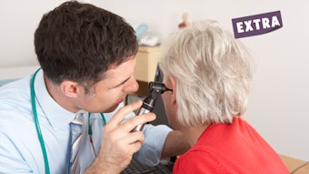
On physical exam, the patient’s vital signs and body mass index (BMI) should be noted, as patients with increased BMI are at increased risk for increased intracranial hypertension. Examination with binocular microscopy should be performed, with careful attention to note any relevant abnormalities, such as a middle ear effusion, tympanic membrane perforations, and middle ear tumors (including glomus tympanicum and glomus jugulare) (Figure 1).
 Figure 1. Evaluation of pulsatile tinnitus.
* Refer to neurotology if cerebrospinal fluid leak is suspected.
** Magnetic resonance angiography and venography can be considered if patient cannot tolerate CT contrast.
Abbreviations: BMI, body mass index; CT; computed tomography; CTA, CT angiogram; CTV, CT venogram; hCG; human chorionic gonadotropin; Hgb, hemoglobin; LP, lumbar puncture; MRI, magnetic resonance imaging; MEE, middle ear effusion; n, nerve; neurotol; neurotology; ophthalm, ophthalmology; TB, temporal bone; TSH, thyroid-stimulating hormone; vasc; vascular.
Periauricular, postauricular, and cervical auscultation can be informative to evaluate for the presence of vascular bruits, which suggest an arterial cause for the patient’s symptoms. In addition to a complete head and neck evaluation, patients who have symptoms at the time of evaluation may undergo gentle jugular vein compression on the side of the symptom and asked to report on whether the sound is affected by this compression. (If bilateral symptoms are present, compression should be performed one side at a time.) An increase in subjective symptomatology with jugular venous compression suggests an underlying venous etiology. A complete audiologic evaluation, including pure-tone and speech audiometry and tympanometry should be obtained with close attention to air-bone gaps and tympanometry findings. Typically, acoustic reflexes should also be performed during these evaluations to aid in the diagnosis of canal dehiscence syndrome.
Some patients may report intermittent symptoms or symptoms of short duration. In such cases, providers may opt to offer an initial period of observation with no further work-up. However, for patients with constant pulsatile tinnitus or persistent symptoms, additional evaluation is recommended with testing choices based on the patient’s initial presentation, medical history, physical examination findings, and audiologic evaluation (Figure 1). This additional evaluation may include laboratory work-up, imaging studies, and subspecialty referrals.
Initial diagnostic studies should be directed by the patient’s history and any notable findings from physical exam. For instance, if the patient’s clinical evaluation is suggestive of a middle ear neoplasm or cerebrospinal fluid (CSF) middle ear effusion, initial evaluation may begin with CT temporal bone. Exam findings suggestive of a vascular origin (bruits and thrills) may prompt concurrent vascular imaging.1,2,3 However, in many cases, the clinical evaluation may not be revealing, and clinicians are left to determine whether to order individual studies sequentially or to order a large battery of tests at once, each with uncertain diagnostic value to that specific patient. In these cases, clinical decision making can be directed by a combination of patient risk factors, guidance from the literature, and practice environment (Figure 1).
Laboratory evaluation with complete blood count and thyroid stimulating hormone (TSH) may assist in the evaluation of anemia and hyperthyroidism. Pregnancy testing may also be indicated. Potential radiographic studies include carotid duplex sonography (or carotid dopplers), high-resolution computed tomography (CT) of the temporal bone without contrast, CT or MR angiography and venography, and contrast-enhanced magnetic resonance imaging (MRI) of the brain and internal auditory canals. Potential consultations include neuro-ophthalmology (for retinal exam and assessment of papilledema in the evaluation of possible IIH), neurology (for lumbar puncture and opening pressure evaluation, again for IIH), and neurosurgery or interventional radiology (for cerebral angiography).
There is no universal consensus on what constitutes the “best” initial radiographic study in the work-up of pulsatile tinnitus. The literature on pulsatile tinnitus is heterogeneous. Reports vary widely due to a combination of differences in study design and modalities of interest, with diagnostic yields ranging from 24%–97% across all modalities. In a recent systematic review and meta-analysis, diagnostic yield varied based on imaging modality, with a low of 21% (95% confidence interval [CI]: 12%–35%) in carotid duplex sonography (CDS) to a high of 86% (CI: 80%–90%) for computed tomographic angiography (CTA). Studies evaluating a multimodal test battery resulted in a yield of 78% (CI: 62%–88%). Some providers may thus consider ordering a CT angiogram of the head for patients who present with pulsatile tinnitus and otherwise have no discernible differentiating factors in their history or on physical exam.5-6 Others have advocated for concurrent CT angiogram and venogram in the assessment of patients with pulsatile tinnitus and a normal otoscopic exam.7 Finally, it is important to note that availability and access to various imaging modalities and subspecialty providers may vary based on practice environment. For some providers, it may be sensible to order the studies most readily available to them or most accurate at their institution before referring the patient to a subspecialist.
When a diagnostic entity is identified on imaging, the next steps should be guided by that diagnosis. Medical issues, including anemia and thyroid disorders, should be treated in conjunction with the patient’s primary care team. Middle ear effusion, glomus tumors, canal dehiscence syndrome, sigmoid sinus dehiscence, and jugular bulb dehiscence are all diagnostic entities that warrant consideration for surgical intervention with an otolaryngologist, otologist, or neurotologist. If the patient’s complete evaluation is negative, the patient may be referred to a neurovascular interventionalist to discuss the risks and benefits of a cerebral angiogram, which may be valuable for the definitive evaluation of intracranial entities including venous sinus stenosis or dural arteriovenous fistula and can increase the rate of definitive diagnosis of underlying etiology >20% after prior non-diagnostic, non-invasive imaging.9
References
- Mattox DE, & Hudgins P (2008). Algorithm for evaluation of pulsatile tinnitus. Acta Oto-laryngologica, 128(4), 427-431.
- Cosetti M, Roehm PC. “Chapter 161: Tinnitus and Hyperacusis,” in Johnson JT and Rosen C. (eds). Bailey’s Otolaryngology—Head & Neck Surgery, Sixth Edition, Wolters Kluwer, Philadelphia PA USA, 2022.
- Tsai LK, Yeh SJ., Tang SC, Hsieh YL, Chen YA, Liu HM, Jeng JS (2016). Validity of carotid duplex sonography in screening for intracranial dural arteriovenous fistula among patients with pulsatile tinnitus. Ultrasound Med Biology, 42(2), 407-412.
- Stevens SM, Rizk HG, Golnik K, Andaluz N, Samy RN, Meyer TA, Lambert PR (2018). Idiopathic intracranial hypertension: contemporary review and implications for the otolaryngologist. Laryngoscope, 128(1), 248-256.
- Cao AC, Hwa TP, Cavarocchi C, Quimby A, Eliades SJ, Ruckenstein MJ, Bigelow DC, Coudhri OA, Brant JA (2023). Diagnostic yield and utility of radiographic imaging in the evaluation of pulsatile tinnitus: A systematic review. Otology & Neurotology Open, 3(2), e030.
- Mohseni M, Asghari A, Daneshi A, Jalessi M, Rostami, S, Nasoori Y. (2018). Comparison of brain CT angiography/venography and temporal bone HRCT scan findings in patients with subjective pulsatile tinnitus in affected side and unaffected side. Biomed Res Therapy, 5(7), 2455-2465.
- Mundada P, Singh A, Lingam RK. (2015). CT arteriography and venography in the evaluation of pulsatile tinnitus with normal otoscopic examination. Laryngoscope, 125(4), 979-984
- Ahsan S F, Seidman M, Yaremchuk K. (2015). What is the best imaging modality in evaluating patients with unilateral pulsatile tinnitus? Laryngoscope, 125(2), 284-285.
- Tao AJ, Parikh NS, Patsalides A. The role of noninvasive imaging in the diagnostic workup for pulsatile tinnitus. Neuroradiol J 35(2): 220-225


