Impact of Chevalier Jackson and Gosta Dohlman on Endoscopic Surgical Therapy for Zenker’s Diverticulum
Alexander T. Hillel, MD Zenker’s diverticulum, or hypopharyngeal diverticulum, develops in a triangular area of weakness between the oblique muscle fibers of the inferior pharyngeal constrictor muscle and the horizontally oriented fibers of the cricopharyngeus muscle. Its location at the interface of the pharynx, neck, and mediastinum makes surgical access difficult and risks severe morbidity. The history of endoscopic treatment of Zenker’s diverticulum demonstrates the process of transition to new surgical therapies based on the sequential efforts of many pioneers. Two otolaryngologists of great renown, Chevalier Jackson and Gosta Dohlman, were critical in advancing the surgical technique and reducing morbidity in the endoscopic treatment of Zenker’s diverticulum. Jackson proposed an esophagoscope-assisted, one-stage, transcervical diverticulectomy with the aim of decreasing morbidity and improving recovery time as compared to the two-stage operation. Jackson’s use of contemporary endoscopic technology advanced the surgical treatment of Zenker’s diverticulum, lowering morbidity in a variety of ways. First, the esophagoscope emptied the diverticulum’s contents, which decreased the risk of aspiration pneumonia and mediastinitis. Second, its distal illumination facilitated identification of the diverticular sac, which in turn cut operative time. Jackson’s use of the esophagoscope represented a vital step in evolution of surgical treatment for Zenker’s diverticulum from an external diverticulectomy to the endoscopic esophagodiverticulostomy. Dohlman attributed cricopharyngeal spasm as the key cause of hypopharyngeal diverticulum, after comparing barium swallow studies of Zenker’s patients to those of controls. His utilization of the cricopharyngeal myotomy directly addressed this key component of disease pathogenesis. In 1960, Dohlman published his case series of 100 patients treated with the endoscopic esophagodiverticulostomy. He reported no cases of low morbidity and recurrence rate, along with much more rapid recovery times, supporting the role of cricopharyngeal myotomy as a key step in the evolution of endoscopic esophago-diverticulotomy. The great leap forward in the general acceptance of endolaryngeal repair of Zenker’s diverticulum can be attributed to the endoscopic stapler, introduced separately in 1993 by Martin-Hirsch et al. and Collard et al. The ability to simultaneously divide and staple the mucosal edges relieved concern about suture-less division of the esophagodiverticular wall. More importantly, the results achieved with the endoscopic stapler technique were more easily reproduced by other otolaryngologists, resulting in more patients treated endoscopically. Endoscopic stapler-assisted diverticulostomy now represents the first line surgical treatment for Zenker’s diverticulum, due to reduced morbidity and shortened operative and recovery times compared with external approaches. *Abridged from: Evolution of endoscopic surgical therapy for Zenker’s Diverticulum. Laryngoscope 2009; 119:39-44.)
Alexander T. Hillel, MD
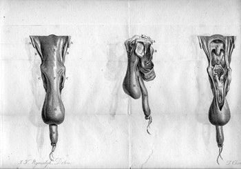 First known published drawings of Zenker’s diverticulum from an autopsy performed by Mr. Abraham Ludlow, surgeon, at Bristol, England in 1764. A) Anterior; B) lateral; and C) open anterior views. (Reproduced with permission from the Institute of the History of Medicine, Johns Hopkins University.)
Zenker’s diverticulum, or hypopharyngeal diverticulum, develops in a triangular area of weakness between the oblique muscle fibers of the inferior pharyngeal constrictor muscle and the horizontally oriented fibers of the cricopharyngeus muscle. Its location at the interface of the pharynx, neck, and mediastinum makes surgical access difficult and risks severe morbidity.
The history of endoscopic treatment of Zenker’s diverticulum demonstrates the process of transition to new surgical therapies based on the sequential efforts of many pioneers. Two otolaryngologists of great renown, Chevalier Jackson and Gosta Dohlman, were critical in advancing the surgical technique and reducing morbidity in the endoscopic treatment of Zenker’s diverticulum.
Jackson proposed an esophagoscope-assisted, one-stage, transcervical diverticulectomy with the aim of decreasing morbidity and improving recovery time as compared to the two-stage operation. Jackson’s use of contemporary endoscopic technology advanced the surgical treatment of Zenker’s diverticulum, lowering morbidity in a variety of ways.
First, the esophagoscope emptied the diverticulum’s contents, which decreased the risk of aspiration pneumonia and mediastinitis. Second, its distal illumination facilitated identification of the diverticular sac, which in turn cut operative time. Jackson’s use of the esophagoscope represented a vital step in evolution of surgical treatment for Zenker’s diverticulum from an external diverticulectomy to the endoscopic esophagodiverticulostomy.
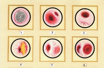 Endoscopic views as sketched by Chevalier Jackson: 1) Zenker’s diverticulum with food bolus; 2) Zenker’s diverticulum following endoscopic removal of the food bolus; and 3) esophagus following diverticulectomy. (Reprinted from Bronchoesophagology and endoscopic plates illustrated by Chevalier Jackson.)
Dohlman attributed cricopharyngeal spasm as the key cause of hypopharyngeal diverticulum, after comparing barium swallow studies of Zenker’s patients to those of controls. His utilization of the cricopharyngeal myotomy directly addressed this key component of disease pathogenesis.
In 1960, Dohlman published his case series of 100 patients treated with the endoscopic esophagodiverticulostomy. He reported no cases of low morbidity and recurrence rate, along with much more rapid recovery times, supporting the role of cricopharyngeal myotomy as a key step in the evolution of endoscopic esophago-diverticulotomy.
The great leap forward in the general acceptance of endolaryngeal repair of Zenker’s diverticulum can be attributed to the endoscopic stapler, introduced separately in 1993 by Martin-Hirsch et al. and Collard et al. The ability to simultaneously divide and staple the mucosal edges relieved concern about suture-less division of the esophagodiverticular wall.
More importantly, the results achieved with the endoscopic stapler technique were more easily reproduced by other otolaryngologists, resulting in more patients treated endoscopically. Endoscopic stapler-assisted diverticulostomy now represents the first line surgical treatment for Zenker’s diverticulum, due to reduced morbidity and shortened operative and recovery times compared with external approaches.
*Abridged from: Evolution of endoscopic surgical therapy for Zenker’s Diverticulum. Laryngoscope 2009; 119:39-44.)
