Innovations in Wound Care for Otolaryngologists
Advances in cell-derived therapeutics, smart-device technologies, and biomaterials are poised to transform skin repair.
Rodrigo Martinez-Monedero, MD, PhD, and Ramesh Balasundari, PhD, on behalf of the Plastic and Reconstructive Surgery Committee
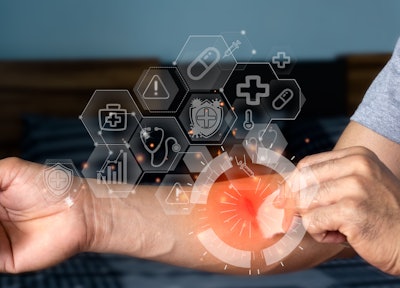
Wound healing is a complex, multistage process involving several cell types. When skin is wounded, its stem cells must respond rapidly to restore the compromised barrier and repair tissue damage. Wound healing involves three overlapping phases: inflammation, tissue formation and tissue remodeling. Inflammation occurs immediately after injury. Following platelet aggregation, various leukocyte lineages, including neutrophils, macrophages, mast cells, and T cells are recruited to the wound site. In addition to clearing dead cells and fighting against infections, these leukocytes secrete cytokines and growth factors such as TGF-βs, IGFs, and FGFs that promote angiogenesis, migration and proliferation of keratinocytes and dermal fibroblasts, synthesis of extracellular matrices (ECMs), and sometimes generation of new hair follicles in the process of epidermal regeneration.
During tissue formation, granulation tissue, consisting of newly formed blood vessels, macrophages, and fibroblasts, begins to cover the wound. Epidermal cells then migrate over the granulation tissue to re-epithelialize the wound. Adipocyte precursor cells are also activated at this stage to generate mature adipocytes important for fibroblast recruitment. During tissue remodeling, the epidermis and dermal fibroblasts deposit new ECM proteins to strengthen the repaired tissue.
After years of basic research, scientists have learned that wound healing differs between animals and humans due to variations in healing rate, mechanisms, skin structure, and immune responses. Humans heal slower, primarily through granulation and re-epithelization, whereas rodents heal more quickly by contraction due to the panniculus carnosus muscle. A key difference in skin includes hair follicles, which are higher in small animals. The immune system also plays a more complex role in human scarring, with animal models often lacking certain immune components relevant to human conditions.1,2 All these findings make translational research challenging because no single animal model perfectly replicates all aspects of human wound healing. To address these limitations, researchers often use multiple animal models, each tailored to a specific biological aspect of wound healing relevant to humans.
With the sequencing of the human genome and advances in DNA sequencing technology, it has become possible to see the whole picture of wound healing. This transforms our understanding of the disease and healing process.
Following full-thickness wounds, cells from both the hair follicles and the interfolicullar epithelium (IFE) migrate into the site of damage. Very few hair follicle stem cell-derived cells persist within the re-epithelialized epidermis of a full-thickness wound. Overall, the data on full-thickness wounds have led to the now widely held view that input from hair follicles plays relatively minor roles, whereas IFE plays a major role in wound re-epithelialization.
Although the specialized cells in the skin have characteristic patterns of gene expression, each cell is capable of altering its pattern of gene expression in response to extracellular cues. Procedures with regenerative strategy scaffolds, growth-factor delivery, cell/exosome therapies, and bioprinting are bringing new options for epithelium and mucosal healing. This change in the expression of the genes of epithelial cells by external signals can have a strong benefit for our patients.
The goal of this article is to summarize the most important recent advances, highlighting translational progress and sketching near-term future directions likely to affect our clinical practice.
Multifunctional and “Smart” Wound Dressings: Making the Dressing an Active Therapy
Traditional dressings may be replaced by multifunctional systems that actively modulate the wound environment. These “smart dressings” integrate antimicrobial agents, controlled-release growth factors, sensors (pH, oxygen, temperature), and stimuli-responsive polymers to deliver payloads on demand.3 Work over the last three to five years has shown improved re-epithelialization, reduced infection, and more regulated inflammation when dressings combine bioactive agents with advanced polymer matrices.
The ideal wound care technology would:
- create a moist, clean, and warm environment
- protect the wound bed from mechanical trauma and bacterial infiltrations
- modulate exudate level
- allow for gas exchange
- promote thermal insulation
- be non-toxic and non-allergenic
- deliver therapeutic compounds essential for healing with an optimal temporal profile.
This strategy is particularly attractive in our otolaryngology clinical practice where small, delicate mucosal or epithelial (e.g., facial skin, tympanic membrane, nasal septum) defects benefit from localized, minimally invasive therapy rather than grafts or flaps. Reviews have been published of multifunctional dressings on the healing epithelium that consolidate the evidence for the clinical utility and promotes an ongoing device development.
Smart wound dressings, while highly innovative, have certain limitations in managing complex wounds. They may not be appropriate for all wound types, particularly those that are extensively infected or large, which often necessitate more intensive, hands-on clinical care. Remote wound monitoring technologies, leveraging smartphone applications and telehealth platforms, enable continuous assessment of wound healing in home settings. These systems provide real-time data to healthcare providers, facilitating early intervention and reducing the need for frequent in-person visits, thereby improving patient outcomes.
Advances in artificial intelligence-driven analytics, three-dimensional wound modeling, and sensor-embedded dressings now allow objective evaluation of wound parameters such as size, tissue composition, and exudate characteristics. This facilitates timely modification of treatment plans and promotes greater patient engagement in self-care. This digital integration has proven particularly beneficial for elderly or rural populations, while also alleviating clinical workload.
However, challenges remain in the widespread adoption of smart wound technologies. The incorporation of advanced materials and electronic components increases production costs, creating financial barriers for both patients and healthcare systems. Additionally, obtaining regulatory approval from the U.S. Food and Drug Administration (FDA) for novel smart bandage technologies is a complex and protracted process. Although many of these devices demonstrate strong potential, most remain in the research and development stage, requiring further validation to establish their safety, efficacy, and long-term reliability. Ensuring accessibility also depends on securing insurance and Medicare reimbursement, which remains a significant hurdle to clinical translation and broad public use.
Cell-derived Therapies: Paracrine Medicine for Tissue Repair
Stem cell-derived therapy with nano-fat uses a process to refine the patient’s own adipose tissue into a concentrated solution of adipose-derived stem cells (ADSCs)4, growth factors, and free lipids, which is then injected to promote tissue regeneration and repair. This minimally invasive regenerative treatment is used to enhance skin health, promote wound healing, restore lost volume, and address issues like skin inflammation and aging. Results are gradual, appearing over weeks and months as the body responds to the injected cells.5
The reparative response induced by the ADSCs leads to improved tissue remodeling, new blood vessel growth (angiogenesis), and immune modulation. In the area of skin rejuvenation, ADSCs improves skin quality, increases dermal cellularity, and boosts collagen and elastic fiber density. By using the body’s own cells, the therapy leverages the body’s natural healing mechanisms for soft tissue repair.
Nano-fat therapy has demonstrated significant potential in enhancing skin wound healing by promoting neovascularization, modulating inflammation, and facilitating collagen deposition. In the context of scar management, nano-fat injections have been effective in improving both hypertrophic and atrophic scars by enhancing tissue elasticity and supporting regeneration of the surrounding skin.6
A key advantage of nano-fat is the use of autologous adipose tissue, which minimizes the risk of immune rejection and provides an easily accessible source of regenerative cells. The procedure is minimally invasive and utilizes small cannulas, which contributes to reduced patient morbidity and faster recovery. Compared with other stem cell-based therapies, nano-fat harvesting and processing can be more cost-effective, as it typically does not require specialized culture systems or prolonged ex vivo expansion, making it a practical and efficient option for regenerative interventions.
Scaffolds, Electrospinning, and 3D/Bioprinting: Structural Templates for Regeneration
Scaffolds created via electrospinning and 3D/bioprinting serve as structural templates for epithelial regeneration by mimicking the extracellular matrix (ECM), providing a 3D microenvironment that guides cell behavior.7 Electrospinning produces nanofibrous mats with a fine, ECM-like architecture, while 3D printing allows for complex, customized macro-structures and shapes with high precision. Combining these techniques can overcome the limitations of each, producing intricate, 3D scaffolds with both the detailed micro-structure of electrospun fibers and the controlled macro-architecture of 3D printing to promote tissue regeneration.
There is a significant focus on applying these scaffolds to challenging wounds by incorporating features that reduce inflammation, remove reactive oxygen species (ROS), and promote new blood vessel formation (angiogenesis). Researchers are exploring 4D printing (3D printing plus time) to create dynamic scaffolds that can respond to stimuli and change their properties over time. While promising, further research and in vivo validation are needed to optimize these complex composite scaffolds and move them toward clinical application and regulatory approval.
Remote wound monitoring uses smartphones apps, smart bandages, and telehealth platforms to track wound healing at home, providing real time data to healthcare providers and improving patient outcomes through early intervention and reduced in person visits. Technologies like AI-powdered analysis, 3D wound modelling, and embedded sensors offer objective assessment of wound size, tissue type, and exudate, enabling timely adjustments to treatments plans and better self-care for patients. This approach increases patient care, especially for rural or elderly populations, while also benefiting clinicians by reducing their caseload.8
Practical Recommendations for Clinicians
Practical recommendations for clinicians include:
- Monitor ongoing clinical trials and device approvals for wound healing.
- Engage multidisciplinary teams (biomaterials engineers, cell therapy units, regulatory specialists) before offering advanced biologic therapies to patients.
- When feasible, minimize surgical trauma and apply precision tools and image guidance to improve wound-healing environments and promote healing using a synergistic strategy for regenerative therapy.
Otolaryngology wound care is entering an era where dressings and grafts do more than cover defects; they actively instruct tissue repair. Smart dressings, exosome therapeutics, electro-spun scaffolds, and patient-specific 3D-printed implants form a convergent toolkit that promises faster, more durable healing with less donor morbidity. Translational and regulatory work remains substantial, but early clinical data supports cautious optimism. Over the next three to seven years, clinicians should expect increasing availability of off-the-shelf regenerative materials and adjunctive biologics, paired with improvements in the monitoring of wound healing staging that together will reshape wound management.
References
- Cirka H, Nguyen TT. Comparative analysis of animal models in wound healing research and the utility for humanized mice models. Adv Wound Care (New Rochelle). 2025;14(9):479-512. doi:10.1089/wound.2024.0082
- Dabiri G, Damstetter E, Phillips T. Choosing a wound dressing based on common wound characteristics. Adv Wound Care (New Rochelle). 2016;5(1):32-41. doi:10.1089/wound.2014.0586
- Rani Raju N, Silina E, Stupin V, Manturova N, Chidambaram SB, Achar RR. Multifunctional and smart wound dressings: a review on recent research advancements in skin regenerative medicine. Pharmaceutics. 2022;14(8):1574. doi:10.3390/pharmaceutics14081574
- Sharma S, Muthu S, Jeyaraman M, Ranjan R, Jha SK. Translational products of adipose tissue-derived mesenchymal stem cells: bench to bedside applications. World J Stem Cells. 2021;13(10):1360-1381. doi:10.4252/wjsc.v13.i10.1360
- Ha DH, Kim HK, Lee J, et al. Mesenchymal stem/stromal cell-derived exosomes for immunomodulatory therapeutics and skin regeneration. Cells. 2020;9(5):1157. doi:10.3390/cells9051157
- Hsu, YC., Li, L. & Fuchs, E. Emerging interactions between skin stem cells and their niches. Nat Med 20, 847–856 (2014). https://doi.org/10.1038/nm.364
- Su Y, Toftdal MS, Le Friec A, Dong M, Han X, Chen M. 3D electrospun synthetic extracellular matrix for tissue regeneration. Small Sci. 2021;1(7):2100003. doi:10.1002/smsc.202100003
- Maxant G, Pastrav M, Gogeneata I, Bajcz C, Bertaux AC. Clinical and medico-economic benefits of remote monitoring of chronic wounds. Int Wound J. 2025;22(3):e70140. doi:10.1111/iwj.70140
