Transition to In-office Treatments: Head and Neck
Advances in technology have propelled the field of in-office diagnostics and minimally invasive surgery and allowed for transition to in-office treatment in head and neck surgery. This decreases lost time from work and costs for patients and allows for more speedy diagnosis and recovery from procedures.
Submitted on behalf of the American Head & Neck Society: M. Boyd Gillespie, MD, MSc, Andrew J. McWhorter, MD, and Cherie-Ann O. Nathan, MD
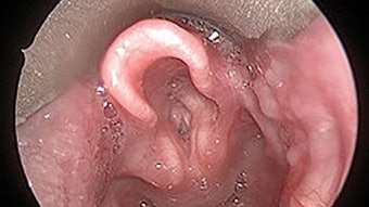
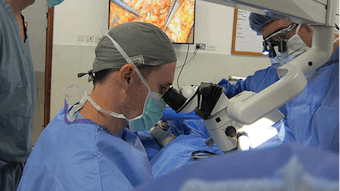
 Figure 1: Photo of Dr. Gillespie’s faculty, Dr. Sandra Stinnett, using office-based laser to treat vocal cord dysplasia.
Advances in technology have propelled the field of in-office diagnostics and minimally invasive surgery and allowed for transition to in-office treatment in head and neck surgery (Figure 1). This decreases lost time from work and costs for patients and allows for more speedy diagnosis and recovery from procedures. The advanced imaging associated with distal chip scopes and minimization of scope caliber while maintaining a working channel has created the opportunity for laryngeal biopsy and treatment of premalignant vocal fold disease with in-office laser treatment with both KTP and CO2 wavelength fiber-based lasers. These procedures previously done in the operating room can safely and cost-effectively be performed while avoiding general anesthesia.
In patients who require a secondary puncture for a trachea-esophageal voice prosthesis, the use of transnasal esophagoscopy combined with multiple described puncture techniques from Seldinger to open are now being utilized in the office. The upright awake patient is sometimes easier to puncture with better visualization. The positioning of the prosthesis as well as having the patient clearing secretions with swallowing and opening the esophagus with swallowing facilitate placement.
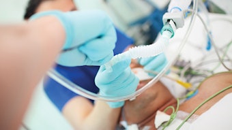
 Figure 2: Photo of an ultrasound needle aspiration of a plunging ranula.
Office-based ultrasonography is becoming an increasingly important tool within head and neck surgery practices. Ultrasonography allows real-time, on-site imaging of a variety of lesions in the thyroid, salivary glands, and lateral neck. Advantages of the technique include efficiency, ability to perform image-guided fine-needle aspiration (FNA) (Figure 2), ability to follow lesions over time without concern for excess radiation exposure, lack of need for contrast enhancement, and relative low cost. It provides additional revenue potential for offices that integrate the findings of U.S. examination within their electronic medical records (EMRs). More recently, several academic head and neck practices have combined office-based ultrasonography with radiofrequency ablation of symptomatic, compressive thyroid nodules not suspicious for cancer following adequate fine-needle aspiration. By applying a radiofrequency probe via percutaneous technique under ultrasound guidance, surgeons are able to achieve up to 50%-80% volume reduction without surgery in select symptomatic nodules. The slower adoption of office-based ultrasonography in the U.S. compared to Europe is likely due to lack of exposure to the technique during residency training, therefore appropriate coursework and ongoing medical education are required for its successful application.
Sialendoscopy (salivary endoscopy) is transitioning from the operating room to the office in practices that have gained experience with the technique. Office-based sialendoscopy allows both diagnosis and management of various obstructive (stones, scar) and inflammatory (chronic sialadenitis, Sjogren’s) conditions of the salivary gland in cooperative adult patients. The technique is most applicable to smaller stones (< 4mm), ductal scars that can be dilated with the tip of the scope or guidewire/bougie system, or hydrostatic dilation and lavage of symptomatic, chronically inflamed glands. Due to the fragility and expense of the scopes, office staff require focused training on scope care and sterilization.
With the COVID-19 pandemic, salivary endoscopy and in particular TEPs and laryngeal in-office laser procedures are high risk aerosolizing procedures and protocols are rapidly evolving. COVID-19 testing prior to the procedure, negative pressure office-based rooms, turn over time between patients in order to allow time for disinfection, use of PPEs, and a number of other measures need to be considered prior to proceeding