Otologic Anatomical Advancement in 18th Century England—Or The Lack of: The Case of Dr. William Cheselden
C. Eduardo Corrales, MD Albert Mudry, MD, PhD At the turn of the 18th century, no suitable atlas of anatomy existed in the English language. In 1713, William Cheselden (1688-1752) published the English manual of anatomy entitled The Anatomy of the Human Body. Contrary to the custom of his day, which preferred Latin (Contugno and Scarpa) or French (Guyot and Petit), Cheselden composed the entire book in English. A unique work, it spanned 93 years with 15 editions including one German edition and three American editions up to 1806. A huge success, the compendium became the preeminent anatomical reference textbook in English-speaking countries. It is largely an anatomical textbook filled with surgical techniques. Cheselden dedicated one chapter to the ear, describing its different anatomical features. In the first edition, Cheselden notably mentions four middle ear ossicles: malleolus, incus, stapes, and officulum quartum; four auditory muscles: externus tympani, obliquus, internus, and stapidis; a description of a tympanic membrane with a small opening; and hearing through the Eustachian tube. In the second edition, he modified the nomenclature of the malleolus to malleus and the officulum to orbicular ossicle, and named the auditory muscles obliquus internus or trochlearis, external oblique, external tympanic, and stapedial. He demonstrated bone conduction through the teeth and discussed the opportunity to perform a myringotomy to improve hearing, which he ultimately performed it on dogs in 1722. He added a “valve” covering the aperture of the tympanic membrane in the third edition. Virtually no modifications to the ear chapter appeared in subsequent editions. Especially interesting are his later editors and publishers in charge of his book. This article primarily demonstrates that Cheselden and his subsequent editors did not critically analyze the otologic knowledge of their time, emphasized by the lack of description of most otologic advances made in the 18th century. Key otologic advances included: A detailed description of the cochlear scalae (Valsalva, 1704); Catheterization of the Eustachian tube through the mouth (Guyot, 1724); The first surgical opening of the mastoid (Petit, 1774); Catheterization of the Eustachian tube through the nose (Cleland, 1744); The first artificial perforation of the tympanic membrane (Bresson, 1748); A description of inner ear fluid (Cotugno, 1760); The inner ear filled with two different fluids (Scarpa, 1789); and A description of the membranous labyrinth (Scarpa, 1795). Possible explanations include: Most treatises of the time were published in French, Italian, or Latin; virtually all otologic advances were published in non-English languages. His treatise was largely a general surgery reference textbook and otology was not a formal surgical specialty. He may have been exposed to secondary references such as Duverney’s Treatise of the Organ of Hearing (1683). Most otologic advances were made in laboratories that combined anatomists, physiologists, and pathologists, and this combination of scientists was mainly to be found in France, Germany, and Italy. Lack of interest of Cheselden and subsequent editors in otologic anatomy may be a reason for lack of otologic advances described in his treatise. Cheselden was undoubtedly one of England’s greatest surgeon-scientists in the 18th century. He was an innovator, skilled politician, and medical illustrator. His compendium spanned 93 years as the anatomical reference book in English-speaking countries. Interested in Our History? Join or renew your membership in the Otolaryngology Historical Society (OHS)—check the box on your Academy dues renewal or contact museum@entnet.org. Save the date—the OHS annual meeting and reception, 6:30 pm-8:30 pm, September 22, in Orlando, FL. Present a paper at the OHS meeting-contact museum@entnet.org. May 15 is the deadline.
Albert Mudry, MD, PhD
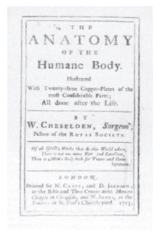
At the turn of the 18th century, no suitable atlas of anatomy existed in the English language. In 1713, William Cheselden (1688-1752) published the English manual of anatomy entitled The Anatomy of the Human Body.
Contrary to the custom of his day, which preferred Latin (Contugno and Scarpa) or French (Guyot and Petit), Cheselden composed the entire book in English. A unique work, it spanned 93 years with 15 editions including one German edition and three American editions up to 1806. A huge success, the compendium became the preeminent anatomical reference textbook in English-speaking countries. It is largely an anatomical textbook filled with surgical techniques.
Cheselden dedicated one chapter to the ear, describing its different anatomical features. In the first edition, Cheselden notably mentions four middle ear ossicles: malleolus, incus, stapes, and officulum quartum; four auditory muscles: externus tympani, obliquus, internus, and stapidis; a description of a tympanic membrane with a small opening; and hearing through the Eustachian tube.
In the second edition, he modified the nomenclature of the malleolus to malleus and the officulum to orbicular ossicle, and named the auditory muscles obliquus internus or trochlearis, external oblique, external tympanic, and stapedial. He demonstrated bone conduction through the teeth and discussed the opportunity to perform a myringotomy to improve hearing, which he ultimately performed it on dogs in 1722.
He added a “valve” covering the aperture of the tympanic membrane in the third edition. Virtually no modifications to the ear chapter appeared in subsequent editions. Especially interesting are his later editors and publishers in charge of his book.
This article primarily demonstrates that Cheselden and his subsequent editors did not critically analyze the otologic knowledge of their time, emphasized by the lack of description of most otologic advances made in the 18th century. Key otologic advances included:
- A detailed description of the cochlear scalae (Valsalva, 1704);
- Catheterization of the Eustachian tube through the mouth (Guyot, 1724);
- The first surgical opening of the mastoid (Petit, 1774);
- Catheterization of the Eustachian tube through the nose (Cleland, 1744);
- The first artificial perforation of the tympanic membrane (Bresson, 1748);
- A description of inner ear fluid (Cotugno, 1760);
- The inner ear filled with two different fluids (Scarpa, 1789); and
- A description of the membranous labyrinth (Scarpa, 1795).
Possible explanations include:
- Most treatises of the time were published in French, Italian, or Latin; virtually all otologic advances were published in non-English languages.
- His treatise was largely a general surgery reference textbook and otology was not a formal surgical specialty.
- He may have been exposed to secondary references such as Duverney’s Treatise of the Organ of Hearing (1683).
- Most otologic advances were made in laboratories that combined anatomists, physiologists, and pathologists, and this combination of scientists was mainly to be found in France, Germany, and Italy.
- Lack of interest of Cheselden and subsequent editors in otologic anatomy may be a reason for lack of otologic advances described in his treatise.
Cheselden was undoubtedly one of England’s greatest surgeon-scientists in the 18th century. He was an innovator, skilled politician, and medical illustrator. His compendium spanned 93 years as the anatomical reference book in English-speaking countries.
 OHS Member, Dr. Lanny Close
Interested in Our History?
- Join or renew your membership in the Otolaryngology Historical Society (OHS)—check the box on your Academy dues renewal or contact museum@entnet.org.
- Save the date—the OHS annual meeting and reception, 6:30 pm-8:30 pm, September 22, in Orlando, FL.
- Present a paper at the OHS meeting-contact museum@entnet.org. May 15 is the deadline.
